


Written by: Eugene Chung
Illustrated by: Sabine Deviche
Illustrated by: Sabine Deviche
show/hide words to know
Note: This activity deals with advanced, potentially sensitive topics. We advise it be reserved for students in middle school or higher grades.
What Is a Brain Tumor?
Your brain is made of billions of cells. These cells communicate with each other so you can see, hear, smell, taste, move, and even think. In very rare cases, cells change in bad ways and mutate. Mutations can make the cells grow uncontrollably so that they form a mass in the brain—a brain tumor. Brain tumors can cause many different symptoms depending on where they are in the brain. Some common symptoms include vomiting, weakness, seizures, headaches, and even changes in personality. They can also make it harder to walk, talk, see, and remember things.
How Can We Treat Brain Tumors?
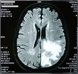
This MRI scan shows a brain with a tumor. Via wikimedia commons(link is external)
Doctors can treat brain tumors many ways. One method is brain surgery. During surgery, a doctor opens the skull and physically cuts out the tumor from the brain. This is called surgical resection. The tumor can also be treated with a chemical mix called chemotherapy. Chemotherapy is toxic and can slow the growth of tumor cells, or kill them. A third option is radiation therapy. In this method, radiation energy is used to kill tumor cells. In many cases, a patient will receive all three treatments (surgery, chemotherapy, and radiation).
Surgery is often the main treatment used. Doctors who do brain surgery are called neurosurgeons and they go through many years of training. Physical removal of a tumor can be hard because tumor tissue can look similar to healthy brain tissue. Doctors want to remove as much of the tumor as possible. However, they have to be very careful because they do not want to hurt or remove any healthy brain.
How do doctors tell the difference between good and bad tissue? Before surgery, doctors will get detailed images of the brain by scanning the head with a CT or MRI machine. These images act like a map for doctors during surgery. Recent medical advances have also allowed doctors to label tumors with fluorescent tags that make the tumor “light up” during surgery. With the help of these fluorescent tags, doctors can visualize tumors. This makes it easier to find and cut out only the bad tissue. Try your hand at new tricks that brain surgeons use. See how much of the tumor you can cut out doing surgery when it is lit up with labels.
What You Need
- Plastic brain mold
- Plastic tumor mold
- Unflavored gelatin
- Water
- Tonic Water
- Black Light
- Tools to remove brain tumor (e.g., plastic knife (“scalpel”), tweezers, spoon straws, etc.)
Before You Begin

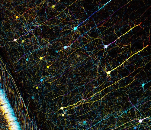
Some of the tumors in this activity will glow and light up under a black light, like these collections of super tiny nano-crystals called quantum dots. Via wikimedia commons(link is external)
Before the surgery, review the different parts of the brain and what each part does. To do so, read through the articles What's in Your Brain? and What's Your Brain Doing? on Ask A Biologist.
It is especially important for brain surgeons to know what different parts of the brain do. If a certain part of the brain is injured, a patient can lose their memory or lose certain functions.
In this activity, you will be trying both old and new techniques that doctors use to remove brain tumors. First, try removing a tumor that does not fluoresce. Next, with the help of a black light, try removing a tumor that lights up.
Procedure
- Read the two stories about brain anatomy and brain function to prepare for the activity. Complete the What’s in Your Brain? (PDF) and What’s Your Brain Doing? (PDF) worksheets.
- Get the two halves of your assigned brains (or two small whole brains). Read the patient description that comes with it. What types of symptoms does the patient have?
- Before you begin your surgery, look at the first brain or brain half. Try to find the tumor. If you find it, what part(s) of the brain is it in?
- After you have found the tumor, try to cut out as much of the tumor as you can. Try not to remove or injure anything that is not the tumor.
- Now with your second brain or brain half, try removing the tumor with the help of the black light. The black light will make the tumor fluoresce.
- After everyone has finished operating, compare the brains or brain halves. Shine the black light on the first brain (half) to see how much tumor you removed. Any remaining tumor will light up. Now look at the second brain (half). In which brain were you able to remove more of the tumor?
Extension: Something went wrong during surgery. Your patient has lost a function. You will be given a scenario describing the patient’s symptoms. Try to figure out what part of the brain was injured during surgery.
View Citation
This activity has a companion article on nerves and the brain. A Nervous Journey takes students on a quick ride from their brain to their toes where they learn about the nervous system and the brain.

Coloring Pages
and Worksheets
Be Part of
Ask A Biologist
By volunteering, or simply sending us feedback on the site. Scientists, teachers, writers, illustrators, and translators are all important to the program. If you are interested in helping with the website we have a Volunteers page to get the process started.