Virtual Bone Lab
We need our bones to walk, run, jump and move, but this is not all they do. Bones are very busy even when you are sleeping at night. They are where blood cells are made and store most of your body's calcium. Bones are also very good at repairing themselves. In this lab you can explore the bones of the human skeleton using our skeleton viewer that can also be played as a game. You can also cut and peel apart a bone to look inside. For a closer look at bones you can use the virtual microscope to inspect their tiniest details.
Skeletal System
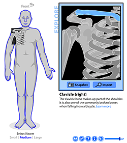
Take a tour of the bones inside the human body with the virtual Skeleton Viewer. In the Explore mode you can select a viewing window and locate the bones of interest. Take a snapshot and then use the inspect icon to click on each bone to see its name and information. The Learn More option links you directly to the companion Wikipedia page for that bone.
Next up, play the skeleton game
Once you master learning the bones, test your knowledge by switching to the Skeleton Game mode to race the clock while you locate and identify bones in the body.
Explore Bone Anatomy
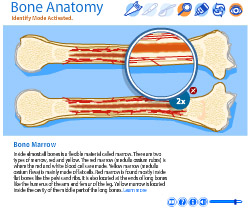
It's easy to look at bones and think of them as dry, dead sticks in your body, but this couldn't be further from the truth. Bones are actually made of active, living cells that are busy growing, repairing themselves, and communicating with other parts of the body.
In our Bone Anatomy Viewer, you can dissect a virtual bone and learn more about the busy world of bones. You can saw, cut away layers, scoop, and zoom into the different parts of a bone. If you want to learn about a bone part, click on the inspect tool to learn what it is and what it does. You can then put it back together with our bone glue.
Virtual Microscope
Take a look at the microscopic world of bone in our virtual bone histology lab. Just pick a microscope slide from below and click on it to view under the virtual microscope. You can zoom in and explore the different parts of the bone. Move around and explore on your own or you can click on the locator buttons to move to a specific part of the bone.
*None of the slide images above are shown at their actual scale. However, when you click and open the virtual microscope, each image has a scale bar that indicates the actual size of the bone sample.
From Micrometer to Millimeter
As you look through the virtual microscope at the slides above, you'll notice a scale bar in the upper corner of each image. You can use this bar to measure how big a part of the image is. As you flip through the slides, you'll notice two different units of measurement, micrometer and millimeter.
1 millimeter (mm) = 1000 micrometer (μm)
To learn more about scale and converting from one unit to another, visit our Matter of Scale article and activity.
Credits: All bone slide images on this page are copyright Professor Jeff Kerr. Used with permission.
Explore Real Bones
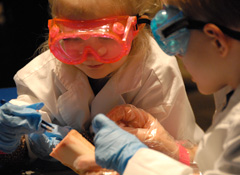
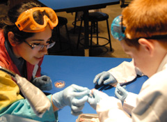
girl and boy kid scientists | lady showing boy bone piece |
Explore bones in person at the Arizona Science Center or schedule a bone lab at your school.
To learn more about field trips, visit the Arizona Science Center Field Trips(link is external) page.
View Citation

Be Part of
Ask A Biologist
By volunteering, or simply sending us feedback on the site. Scientists, teachers, writers, illustrators, and translators are all important to the program. If you are interested in helping with the website we have a Volunteers page to get the process started.


